What is Milia or milialar?

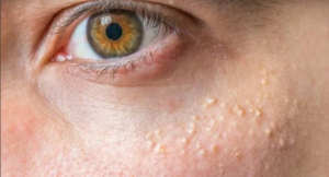
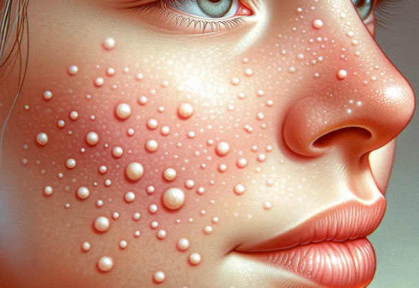
milialar are small, dome-shaped bumps that typically range in size from 1-2 millimeters, about the size of a pinhead. They appear as whitish-yellow, pearly cysts with a firm and smooth texture on the skin surface. The most common location is milia on eyelid and the skin under the eyes, where they resemble tiny pearls embedded under the skin. According to a study, milialar occurs when keratin, a protein found in the skin, hair, and nails, becomes trapped beneath the skin’s surface. While they are often seen in newborns, adults can also develop milialar, often as a result of skin damage.
Fun Fact: Despite the similar-sounding name, milia (singular: milium) or milialar are entirely unrelated to malaria, which is a disease caused by a parasite.
Types of milia or milialar
milialar can be classified into two main types: Primary milialar and Secondary milialar.
Primary Milia or milialar
Primary milialar is directly formed due to the entrapment of keratin within the skin. They are more commonly seen in neonates due to immature sweat ducts. Key characteristics of primary milia or milialar include:
- Small, white-to-yellow cysts.
- Commonly found on the face, especially on the cheeks, nose, and around the eyes.
- Generally asymptomatic.
- Often resolves spontaneously in infants within a few weeks to months.
Secondary Milia or milialar
Secondary milialar, on the other hand, arise as a result of trauma or injury to the skin. They can occur in adults after certain skin conditions or procedures. Key characteristics of secondary milia or milialar include:
- Similar appearance to primary milialar, often seen in areas where an injury or procedure occurred.
- Symptoms might be associated with the underlying cause, such as pain from a burn.
- Duration varies and can persist longer depending on the cause.
- Addressing the underlying cause is crucial, and treatments like manual extraction, laser therapy, or medications may be considered.
What Causes Milia or milialar Development?

milialar form when dead skin cells become trapped under the skin’s surface, resulting in the formation of tiny cysts. While they often appear on the face, especially around the eyes and cheeks, they can also occur elsewhere on the body. Several factors contribute to the development of milia or milialar, although the underlying trigger is not always identifiable. These factors include:
- Genetics: Some individuals have a hereditary predisposition for developing milia, and it often runs in families.
- Sun Exposure: Prolonged sun exposure can damage facial skin over time, increasing the risk of milia.
- Skin Trauma: Injury to the skin, such as cuts, burns, abrasions, and blisters, may lead to the formation of milia during the healing process.
- Certain Medical Conditions: Disorders that cause dry skin and inflammation, like eczema, can increase the risk.
- Medications: Some medications, like steroids, may promote milia as a side effect.
- Heavy Creams and Makeup: Using thick, greasy products can clog pores and cause cysts.
milialar is most common in newborns, with up to 50 percent of infants developing transient milia that typically go away within a few weeks. The role of maternal hormones is believed to contribute to this occurrence. However, persistent milialar affects approximately 2.5% of the general adult population. Women are more frequently affected than men, and milia become more prevalent with age, thought to be caused by age-related changes in skin cell kinetics and decreased skin elasticity.
Development Process
The development of milialar follows a specific process:
- Skin Renewal: As part of its renewal process, the skin naturally sheds dead cells. Sometimes, these cells don’t shed properly.
- Trapped Keratin: The trapped cells then form keratin, which accumulates.
- Formation of Cysts: This accumulation results in the formation of tiny cysts beneath the skin, leading to milia.
Expert Insight: There are several subtypes of milialar, but primary milialar arising spontaneously due to keratin entrapment are most common around the eyelids. Secondary milialar can arise from trauma, burns, blistering, or ophthalmic conditions.
Signs and Symptoms of Milia or milialar

milialar are generally easy to recognize due to their characteristic appearance. They can manifest as:
- Small, pearly white bumps on the eyelids or around the eyes.
- Dome-shaped, smooth bumps resembling pearls under the skin.
- Whitish-yellow or yellowish-white in color.
- May appear singly or in clusters.
- Typically painless and don’t cause itching or irritation.
- Can remain unchanged for weeks to months or disappear on their own.
- Sometimes may secrete a waxy, cheese-like discharge if ruptured.
Tip: If you’re uncertain about any skin condition or have concerns, it’s always advisable to consult a dermatologist for a proper diagnosis and guidance.
Preventive Measures
While it may not be possible to prevent milialar entirely, the following tips can be helpful in reducing the risk of their development:
- Use oil-free, non-comedogenic moisturizers and makeup.
- Avoid heavy, greasy creams and cosmetics near the eyes.
- Cleanse gently and exfoliate the skin regularly to unclog pores.
- Shave carefully using proper technique to avoid injuring the skin.
- Wear sunscreen daily and limit unprotected sun exposure.
- Keep your skin well-hydrated to prevent excess dryness.
- Remove makeup thoroughly before bedtime and discard old makeup.
- Treat any underlying skin conditions like eczema.
- If you are prone to milia, sometimes called millia, consider avoiding intensive facials or chemical peels which may worsen them.
Treatment Options for Milia or milialar
In most cases, milialar does not require treatment, and many resolve spontaneously within weeks to months. However, if the bumps persist or cause distress, several treatment options are available:
- Prescription Retinoid Creams: Creams containing tretinoin, adapalene, or tazarotene can help dry out and slough off the milia.
- Microdermabrasion: This technique uses fine crystals to gently exfoliate the outer skin layers and stimulate healing.
- Chemical Peels: Applying a mild glycolic or salicylic acid solution helps soften and remove the lesions.
- Electrocautery: This involves burning off the milialar with a hyfrecator cauterizing device, with the use of a local anesthetic.
- Manual Extraction: A dermatologist can open the cyst with a sterile needle and squeeze out the contents.
- Cryotherapy: Freezing the bumps with liquid nitrogen to eliminate the lesions.
- Laser Ablation: Using laser energy to destroy the cysts.
- Surgical Removal: In some cases, a dermatologist may opt for surgical removal by cutting open and draining the milia, sometimes requiring stitches.
Important Note: This information is provided for educational purposes, and it is recommended to seek professional advice from a dermatologist before pursuing any treatment.
Conclusion
In conclusion, milialar, the small pearl-like cysts that can appear on the skin, are generally harmless and can be found on the eyelids and around the eyes. While they are more common in newborns, adults can also develop them, often as a result of skin damage. milialar comes in different types, and their development is influenced by various factors, including genetics, sun exposure, skin trauma, medical conditions, medications, and the use of heavy creams and makeup.
If you have milialar and they cause you distress, there are various treatment options available, although many cases resolve naturally. To prevent milia, it’s essential to adopt proper skincare practices, avoid heavy cosmetics, no makeup and protect your skin from excessive sun exposure.
This comprehensive guide has provided you with a detailed understanding of milia, its types, development, signs and symptoms, preventive measures, and treatment options. If you have any concerns about your skin or milialar, consult a dermatologist for professional guidance.


